Tag

Tagged: spine surgery

Sponsored
  • Over the past 2 decades minimally invasive surgery and computer assisted navigation (CAN) systems have significantly changed spine surgery
  • Minimally invasive spine surgery (MISS) has become a significant subspeciality accounting for ~50% of all spine surgeries undertaken in the US
  • Together MISS and CAN systems promise enhanced precision, improved outcomes, and lower costs
  • CAN systems provide surgeons with improved visibility of the operating site, but emit hazardous radiation that can cause cancer
  • Spine surgery appears to be winning the challenge to increase the development of minimally invasive surgery while decreasing harmful radiation in the operating room
  • MISS is positioned to grow and increase its market share but faces some headwinds
 
- Low back pain and the global spine industry - 
 
Minimally invasive spine surgery and computer assisted navigation systems
 
Minimally invasive spine surgery (MISS) requires only a small incision and uses specialized instruments and techniques that minimize cutting and results in minimal damage of body tissue. The technique serves the increasing prevalence of degenerative spinal disorders, attributed to sedentary lifestyles of aging populations, which have helped to drive the growth of a global spinal implants and devices market. In addition to the increased availability of biologics and customizable implants and the refinement of operative techniques, the development of MISS has been supported by advances in imaging and navigation technologies that make surgical targets virtual on a monitor to improve the accuracy and precision of surgical interventions. Today, there is a growing body of research demonstrating MISS’s advantages over the traditional open approach.  However, computer assisted navigation (CAN) systems tend to emit harmful ionizing radiation that can cause cancer. Reducing radiation in the OR while improving the quality of image guidance is expected to fuel further growth of MISS.
 
 In this Commentary

This Commentary focuses on minimally invasive spine surgery and computer assisted navigation systems. Two technologies, which have changed the landscape of modern spine surgery and offer potential benefits for both patients and surgeons. Has MISS reached its market saturation? If not, what will affect the speed and extent of its further adoption? 
 
Minimally invasive and open spine surgery

Over the past 2 decades, MISS has become a significant subspeciality and currently accounts for ~50% of all spine surgeries undertaken in the US. It is positioned to increase its influence over the next decade but faces some headwinds.

As a general principle, it is preferable to intrude as little as possible when carrying out a surgical procedure to minimise damage to surrounding tissue and to speed up recovery time. Many spine procedures that once required invasive operations (open surgery) have been replaced with MISS techniques.

Open spine surgery typically involves relatively long incisions down the back to give the surgeon the best view of, and access to, the anatomy. During such procedures, it is sometimes necessary to cut through and move aside muscles and tendons to reach the affected area, which can cause damage to these tissues and prolong recovery.

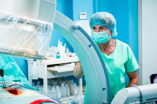
In MISS the surgeon makes a small incision and then inserts a device called a tubular retractor, a stiff, tube-shaped tool that creates a tunnel to the problem area of the spine by gently pushing aside the muscle and soft tissue around the affected area. The surgeon can then put small tools through the tunnel to work on the spine and use a special microscope to view real-time X-ray images of the spine. This approach results in less damage to the muscles and soft tissues that surround the spine, which leads to a more expedited recovery.

MISS has gained popularity both with patients and clinicians and has become increasingly feasible for the management of a range of spinal disorders. Progress has been made in the development of a direct lateral approach [from the side] as well as improvements of tubular retractors. Common spine surgery treatments available through minimally invasive methods include degenerative disc disorders, herniated discs, lumbar spinal stenosis, spinal deformities such as scoliosis, spinal infections, spinal instability including spondylolisthesis, vertebral compression fractures, and spinal tumours. In 2020, MISS procedures accounted for ~50% of all spine surgeries performed in the US, which had increased from ~16% in 2012.

According to David Bell, a consultant neurosurgeon at King’s College Hospital, London, who specialises in complex spine surgery, MISS significantly improves the patient experience by, “reducing the size of the incision and the amount of tissue manipulation . . .  It also minimises post-operative discomfort, cuts infection rates, lessens blood loss and reduces a patient’s recuperation time”. See video below.
 
 
The evidence

There is a growing body of research to support the benefits of MISS, which include: (i) reduced trauma to muscles and soft tissue, (ii) better cosmetic results from smaller incisions, (iii) less blood loss, (iv) reduced risk of infection, (v) faster recovery time and less rehabilitation, (vi) diminished reliance on pain medications, and (vii) reduced hospital stays. A further perceived benefit is the increasing range of MISS undertaken in outpatient settings. Such benefits are likely to fuel the refinement of surgical techniques based on patient outcomes, and lead to the growth of MISS.
 
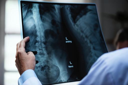
However, not all studies are so positive about the benefits of MISS. A 2017 review of 17 randomized controlled trials, which compared MISS against open procedures for three common disorders, concluded that, “the evidence do not support MISS over open surgery for cervical or lumbar disc herniation”. The study suggests that there were some advantages for transforaminal lumbar interbody fusion (TLIF), [a procedure that melds the front and back sections of the spine through a posterior approach], but “at the cost of higher revision rates, higher readmission rates and more than twice the amount of intraoperative fluoroscopy”. [an imaging technique employed to improve intraoperative visualization of the operating field, which emits hazardous radiation]. The study concludes that, “Regardless of patient indication, MISS exposes the surgeon to significantly more radiation”. 

Two papers published in the January 2020 edition of the Journal of Spine Surgery report on a global survey of 430 surgeons to assess the extent of MISS and the training surgeons receive. The response rate was significant at 67%. 33% of respondents were neurosurgeons, 55% orthopaedic surgeons and 12% were surgeons with other postgraduate training. One research paper concludes that, “endoscopic spinal surgery is now the most commonly performed MISS technique”, and the other suggests that, “very few MISS surgeons are fellowship trained but attend workshops and various meetings suggesting that many of them are self-thought. Orthopaedic surgeons were more likely to implement endoscopic spinal surgery into the routine clinical practice”.
A review of the state of MISS reported in the June 2019 edition of the Journal of Spine Surgery confirms MISS as a significant subspeciality, “evidenced by the large and constantly growing body of literature on this topic”, and driven by “significant advancements in imaging and navigation technologies, refinement of operative techniques, availability of biologics and customizable implants, and most importantly, evidence of feasibility, efficacy, safety and value, compared to traditional approaches as demonstrated by the current literature”.
You might also like:

If spine surgery fails to relieve low back pain why is it increasing?


Unmistakably, over the past two decades, MISS has become increasingly feasible, efficient, and popular. An important question is, how fast is MISS advancing? There is a paucity of research, which addresses this question. However, a global survey of spine surgeons published in the January 2020 edition of the Journal of Spine Surgery provides some insights. Findings suggest there are regional variations in the acceptance and utilization of MISS. The study surveyed 586 spine surgeons in 5 major regions of the world, which yielded 292 usuable responses: a significant response rate of ~50%. 70% of spine surgeons in Asia and South America thought MISS was accepted into mainstream spinal surgery in their practice areas compared to 63% of spine surgeons in North America, 53% in Europe and 50% in Africa & the Middle East. The percentage of spine surgeons that reported using MISS was higher: Asia (97%), Europe and South America (89%), and Africa & the Middle East (88%). Surgeons in North America reported the lowest rate of MISS implementation globally.  
 
Although innovations and techniques in MISS have continued to develop over the past decade, a significant percentage (~50% in the US) of surgeons are understood to use open surgical techniques. Reasons for this include: (i) lack of adequate surgeon training and experience, (ii) the steep learning curve needed for MISS, (iii) inadequate hospital resources and (iv) the patchiness of research on the benefits of MISS. It seems reasonable to suggest that such factors affect the adoption rate of MISS. But perhaps the most significant factor influencing the speed of its adoption will be the rate of development of robotic surgical systems. An understanding of the impact of these factors will help producers hone their strategies and business models.
 
Computer assisted navigation systems

A common therapy to correct spinal disorders is fusion, which melds together two or more vertebrae so that they heal into a single, solid bone. Spinal fusion surgeries use implants of biocompatible materials, such as titanium, as well as rods, plates, screws, and interbody cages and account for the largest segment of the global spine market.
You might also like: 

Low back pain, spine surgery and market shifts

During spinal fusion procedures, pedicle screws are used for spinal fixation, stability, and fusion. The incorporation of such screws into spine surgery in the early 1960s was a significant advance because it offered stability and decreased rates of pseudarthrosis [failure of a fractured bone to heal] compared to previous methods. However, subsequent studies suggested that there was a percentage of pedicle screws inaccurately placed, which could harm adjacent structures and potentially have mechanical, neurological, and vascular consequences for patients.
Image guidance systems were a noteworthy development in spine surgery to reduce the morbidity associated with the mispositioning of pedicle screws and today such systems are used widely. Fluoroscopy, an early guidance method, provided real-time X-ray imaging for guiding interventional procedures, which resulted in more accurate placement of screws, but such systems emitted hazardous ionizing radiation that surgeons, patients, and OR staff were subjected to. Any spine surgery that is visualized with fluoroscopy can involve 10 to 12X higher amounts of radiation from the use of X-rays compared to non-surgical procedures. Compared to a hip surgeon, a spine surgeon can experience 50X more harmful emissions over the course of a professional career and this has been linked to the development of cancers. Reducing radiation exposure is an important challenge.
 
Guided systems and reduced radiation

Newer intraoperative navigation modalities have been found to reduce radiation exposure significantly compared to traditional fluoroscopic guided percutaneous surgical techniques, and have become an important addition in spine surgery. Real-time image guidance, along with continuous computation and scan integration by the navigation system, allows a surgeon to visualize a comprehensive 3D picture of the operating site. Intraoperative computerized tomography (CT) scans [the use of X-rays and a computer to create detailed images of the operating site], together with infrared and other optical guidance technologies have substantially increased the accuracy and precision of spine surgeons to place pedicle screws. 
 
One such enhanced guidance system is ultralow radiation imaging (ULRI) coupled with image enhancement and instrument tracking (IE/IT). This is a new image modifier that allows a computer to show real-time movement of an instrument as it is adjusted, mimicking live fluoroscopy, but without continuous radiation production. Recent research suggests that ULRI-IE/IT systems, “can dramatically reduce radiation output and the number of images acquired and time needed to perform fluoroscopic procedures”. 
 
There are numerous FDA approved advanced CAN systems but let us briefly describe some popular ones. The Airo Mobile Intraoperative CT-based Spinal Navigation system was approved by the FDA in 2013, and developed by Brainlab, a privately held German MedTech company headquartered in Munich. The technology is one of the pioneers of advanced surgical navigation platforms and has many similarities to other CAN systems. It uses a mobile circular scanner attached to the operating table for 360° imaging, and a scanning stereotactic camera, which uses a set of three coordinates for instrument registration. Research published in the July 2018 edition of the Journal of Neurosurgery suggests that the Airo “mobile CT scanner reduced the rate of screw repositioning, which enhanced patient safety and diminished radiation exposure for patients, but it did not improve overall accuracy compared to that of a mobile 3D platform”.
 
Another popular system is Medtronic’s Stealth Station Spine Surgery Imaging and Surgical Navigation with O-arm, a portable imaging device that fits over the surgical table to take images of the operating field. This uses similar technology to Brainlab’s Airo, but opens at 90° to allow for mobilization around the patient. A third system is produced by Ziehm Imaging, another German company, which specializes in the development and manufacture of mobile C-arms [imaging devices that can be used flexibly in operating rooms]. In 2015, the company received FDA approval for the Ziehm Vision FD Vario 3D with NaviPort Integration. This is an intuitive technology, which obtains images via a 190° rotation with a C-arm around the patient and provides surgeons with, “crystal-clear and distortion-free 3D images for maximum intraoperative visualization of anatomical structures”. However, if its reference clamps are moved after the initial registration process, repeat CT scanning is required to re-register the clamps. Stryker’s SpineMask Tracker and SpineMap Software system overcome this problem by gluing its reference trackers to patients.
 
With the widespread use of CAN systems in spine surgery there is an increasing number of studies, which demonstrate the advantages of such technologies. For example, two large meta-analyses suggest that CAN systems significantly increase the accuracy of pedicle screw placement compared to freehand placement. Research also suggests that patients who undergo CAN pedicle screw placement have lower complication rates than those who undergo freehand placement.
 
Notwithstanding, findings of a global survey conducted in 2013 and reported in the September 2019 edition of The Spine Journal suggest that ~78% of surgeons still use two-dimensional fluoroscopy during spine surgery. Despite the improved accuracy and reduced radiation provided by advanced computer-assisted spine navigation systems. This could be associated with costs, prolonged operative times, and their cumbersome nature.
 
Machine-vision image guided surgery system

7D Surgical, a Toronto based company that develops advanced optical technologies, has sought to overcome challenges inherent in traditional CAN systems by developing a machine-vision image guided surgery platform, [FLASH™]. The technology employs a satellite-based global positioning system (GPS), to create a 3D image of a patient’s anatomy, and uses visible light coupled with machine-vision algorithms that eliminate exposure to intraoperative radiation. Other benefits of 7D’s system include its rapid set up time and its minimal workflow disturbance. The fact that its navigation camera is integrated into the surgical light, eliminates the need to stop surgery and position supplemental surgical equipment, thereby allowing for continuous access to the surgical field. Further, and unique to FLASH™, is the fact that its reference clamp can be repositioned, and images re-registered within ~20 seconds. This facilitates seamless clinical applicability and reverses many of the drawbacks of preceding navigation systems. In May 2021, SeaSpine, a Nasdaq traded spine company, announced the acquisition of 7D in a deal valued at US$110m. In July 2021 SeaSpine received FDA approval of 7D’s advanced guidance system for MISS.
 
Takeaways

Over the past two decades, MISS has had a significant impact and established itself as a subspeciality throughout the world. Although it is difficult to calculate, it appears that ~50% of spine surgeries could still be open procedures. This suggests that strategic questions facing producers include whether MISS will expand further, and if so, at what speed. This Commentary suggests some factors, which are likely to impede the adoption rate of MISS. However, perhaps the most significant challenge to MISS is not the prevalence of open surgery, but the rapid rise and adoption of robotic surgical systems. Research published in the January 2020 edition of the Journal of the American Medical Association on the trends in the adoption of robotic surgery concludes, “Hospitals that launched robotic surgery programs had a broad and immediate increase in the use of robotic surgery, which was associated with a decrease in traditional laparoscopic minimally invasive surgery”. Robotic surgical systems in spine surgery is the subject of a forthcoming Commentary.
view in full page
  • People are living longer, the prevalence of age-related degenerative disc disease is increasing and sufferers are more and more turning to spinal implant surgery as a solution
  • As this significantly raises the burden on over-stretched healthcare systems, so is spine surgery increasingly becoming a key target for cost reduction within healthcare systems
  • This intensifies the pressure on manufacturers to innovate and make spinal implants more cost effective

Can 3D printing and the use of new alloys reduce the high costs of producing and marketing spinal implants?
 
On January 8th 2019 surgeons at Joseph Spinea specialist surgery centre based in Tampa Bay Florida, were the first in the US to implant a 3D printed interbody fusion device, which was produced  by Osseus Fusion SystemsThe company uses its proprietary 3D printing technology, also known as additive manufacturing,  to build spinal implants from titanium material that is optimized for bone fusion and biological fixation. In August 2018, a suite of Osseus’s devices received clearance from the US Food and Drug Administration (FDA) for a  range of heights and lordotic (inward spinal curvature) angles, which make them adaptable for a variety of patient anatomies. The interbody fusion devices work by being packed with biomaterials and bone grafts and inserted in between two vertebrae, where they fuse with the spine and work to prevent back pain.
 
In this Commentary
 
This Commentary explores whether 3D printing and the use of new alloys could be an appropriate strategy to help spine companies reduce  their production and sales costs and enhance their market positions. Our suggestions here complement a strategy, described in a previous Commentary, for MedTech companies to develop and implement digital strategies to enhance their go-to-market activities, strengthen the value propositions of products and services and streamline internal processes. The reasons spine companies might consider both strategies are because spinal implant markets, which are segmented  by type of surgery, product and geography, are experiencing significant competitive, regulatory, pricing and technological challenges as well as mounting consumer pressure for improved outcomes; and the business model, which served as an accelerator of their financial success over the past decade is unlikely to be effective over the next decade.
 
3D printing
3D printing is a process, which creates a three-dimensional (3D) object by building successive layers of raw material. Each new layer is attached to the previous one until the object is complete. In the healthcare industry, 3D printing is used in a wide range of applications, such as producing dental crowns and bridges; developing prototypes; and manufacturing surgical guides and hearing aid devices. Increasingly, 3D printing is being used for the production of spinal implants.

 
Spine surgery increasing significantly
 
An estimated US$90bn is spent each year in the US on the diagnosis and management of low back pain (LBP). LBP, caused by age related degenerative disc disease, is one of the most common and widespread public health challenges facing the industrialized world. It is estimated that the condition affects over 80% of the global population and inflicts a heavy and escalating burden on healthcare systems. Also, LBP affects  economies more generally in terms of lost production due to absenteeism, early retirement and the psychosocial impact caused by the withdrawal of otherwise active people from their daily activities. According to the American Association of Neurological Surgeons, more than 65m Americans suffer from LBP annually and the Chicago Institute of Neurosurgery and Neuroresearch suggests that by the age of fifty, 85% of the US population is likely to show evidence of disc degeneration. It is estimated that 10% of all cases of LBP will develop chronic back pain, which is one of the main reasons for people to seek surgical solutions and this significantly raises the burden on over-stretched healthcare systems.
 
Findings of a study published in the March 2019 edition of Spine, entitled, “Trends in Lumbar Fusion Procedure Rates and Associated Hospital Costs for Degenerative Spinal Diseases in the United States 2004 to 2015”, report that the rate of elective lumbar fusion surgeries in the US has increased substantially over the past decade. Such trends are indicative of most advanced industrial societies, which  are changing and ageing, primarily driven by improvements in life expectancy and by a decrease in fertility. This results in people living longer, reaching older ages and having fewer children later in life. Over the next decade, these market drivers are expected to make spine surgery a key target for cost reduction within healthcare systems and this, in turn, is likely to increase pressure on manufacturers of spinal implants to make spine surgery more cost effective.


The first surgery using a 3D printed spinal implant
 
The first surgery to implant a 3D printed interbody fusion device was carried out in China in August 2014, when surgeon Liu Zhongjun from Peking University Hospital successfully implanted an artificial 3D printed vertebra into a 12-year-old bone cancer patient to help him walk again. Liu first removed a tumour located in the second vertebra of the boy's neck before replacing it with the 3D printed implant between the first and third vertebrae to allow him to lift his head. “The customized 3D printed technology made the disc replacement stronger and more convenient than normal procedures”, said Liu.

You might also be interested in:

Age of the aged and low back pain
 
In July 2017, a team of doctors, led by Xiao Jianru, Professor of Orthopaedic Surgery at Shanghai Changzheng Hospital, China, treated a 28-year-old woman with a massive, rare neck tumour, by giving her a 3D printed spine. The patient had to have six consecutive cervical vertebrae replaced because they had been affected by the cancer, which was challenging to treat with chemotherapy. Cervical vertebrae, seven in total, which form your spine column in the neck are the most delicate bones in your body. The patient was discharged from the hospital after the operation. Reports suggest that she was able to walk, but had some difficulties turning her head.
 
First US company to receive FDA approval for 3D printed spinal implants
 
The first US  company to receive a 510(k) FDA approval for a 3D printed spinal implant was 4WEB Medical, in 2012. The company was founded in 2008 and since then has become a leader in 3D printed implant technology. Following FDA clearance, the company launched its proprietary and patented Truss Implant platform, which features a unique open architecture that allows for up to 75% of the implant to be filled with graft material and includes an anterior spine Truss System, a cervical spine Truss System, an osteotomy Truss System and a posterior spine Truss System. In April 2018,  at the annual meeting of the International Society for the Advancement of Spine Surgery (ISASS) 4Web announced that it has surpassed 30,000 implants worldwide of its proprietary Truss Implant Technology.
 
There is a plethora of established MedTech companies entering the 3D printing spinal implant market, which include Stryker, K2M, DePuy Synthes, Camber Spine, CoreLink, Medicrea, Renovis, NuVasive and Zimmer Biomet. With Stryker’s acquisition of K2M and DePuy Synthes’ acquisition of Emerging Implant Technologies GmbH (EIT), both in September 2018, the market for 3D printed spinal implants is positioned to grow rapidly over the next few years.
 
Increasing FDA approvals for 3D printed spinal implants
 
Significantly, spinal implants have become one of the most common cases of the FDA-cleared 3D printed medical devices. For instance, in 2018 Zimmer Biomet received FDA clearance for the company’s first 3D printed titanium spinal implantEIT received FDA approval in 2018 for its 3D printed multilevel cervical cage, which can treat multiple injuries in both the middle and top parts of the spine. Centinel Spine Inc, a US company based in Pennsylvania, which develops, manufactures and markets spinal devices used to treat degenerative disc disease, also received FDA clearance in 2018 for its 3D printed spinal implants called FLX devices, which are titanium fusion implants that work to stabilize vertebrae from the front of the spine in order to increase the healing process for patients.

You might also be interested in:

MedTech must digitize to remain relevant

 
3D printing medical devices market
 
The 3D printing medical devices market is projected to grow at a CAGR of 17.5% and reach US$2bn by 2022. Currently, the market is dominated by North America, followed by Europe, Asia Pacific and the rest of the world. Over the next decade, the Asia Pacific 3D printing medical devices market is expected to grow at the highest CAGR. Emerging markets are attractive for spine companies as they have large patient populations, which are growing fast, rising government healthcare expenditure, vast and rapidly increasing middle classes, rising income levels and rising obesity cases.
One example is India, with a middle class about twice the size of the US population, an economy growing at a rate of 7% year-on-year and a pro-business Prime Minister who has established himself as the country’s most formidable politician in decades and is committed to increasing healthcare spending. According to the World Bank’s March 2018 India Development Update the GDP of India had surpassed that of France and was on track to overtake the UK economy to make India the 5th largest economy in the world. Significantly, India’s GDP per capita has reached US$2,000, which is generally recognised by economists as a “tipping point”: when a country’s economic prospects improve, peoples’ confidence increases, and investment momentum remains at a desirable level for a long period. For instance, when the GDP per capita of China and South Korea reached US$2,000 their respective economies witnessed more than a decade of high growth with an average growth rate of about 10%. India appears to be on the cusp of something similar.
 
3D printing's competitive advantages
 
3D printing, although in its infancy, has the capacity to manufacture products of any complexity anywhere, at any time, which gives it a significant competitive-advantage over traditional manufacturing. Further, 3D printing is cheaper and quicker than traditional production methods because there is less machine, material, labour and inventory costs and less materials' waste. Complex designs can be created as a computer added design (CAD) model and then transformed into a reality in just a few hours. By contrast, traditional manufacturing methods can take weeks or even months to go from the design stage to a prototype and then onto the production process. Also, 3D printing is cost-effective in low production quantities and more environmentally friendly as the place of manufacture can be the same as the place of the product’s application.

The benefits of 3D printing specifically for spinal surgery include; (i) implants can be shaped to custom-fit patients, (ii) porosity and pore size can be personalized to a specific patient’s bone quality, which may improve integration. But perhaps the most significant potential advantage is bioprinting, where cells, growth factors and biomaterials are used to create living tissue.

 
Thinking beyond traditional metals used for spinal implants
 
Some spine companies are complementing their 3D printing endeavours by experimenting with new and stronger alloys. For the past two decades metals used for spinal implants have been mostly composed of cobalt chrome, titanium and stainless steel. The physical properties of these have prevented producers to reduce the size of spinal implants. But this is changing with the introduction of new alloys such as molybdenum-rhenium (MoRe), which is stronger than the traditional metals used for spine implants and has the potential to use less metal to achieve stronger, more durable constructs, while allowing for smaller sized products.

Already, MoRe is used for stents in cardiology and findings of a small animal study presented at the 2018 North American Spine Society meeting in Los Angeles suggested that MoRe is significantly more hydrophilic (having strong affinity to water) and therefore friendlier to bone when compared with cobalt chrome, titanium and stainless steel. This suggests MoRe might provide smaller rods with smaller pedicle screw heads, which decrease the prevalence of protruding, painful hardware in patients with wasting of the body due to severe chronic illness. Further, smaller spinal implants would be beneficial in minimally invasive spine surgery.

Another added benefit of MoRe is that it potentially decreases biofilm formations, which are typically caused by chronic medical device-related infections and allergenicity when compared to the traditional metals used in spine surgery. Bacteria are tougher to kill when they attach to the surface of a spinal implant, even before they form a biofilm. Research findings published in the December 2018 edition of Heliyon draws attention to the prevalence of the  antibiotic-resistant nature of bacterial biofilm infections on implantable medical devices and describes current state-of-the-art therapeutic approaches for preventing and treating biofilms. As the range of materials for spinal implants with improved biocompatibility, biodegradability and load bearing properties increase, so are biofilm infections expected to decrease.

 
Takeaways
 
Spine surgery is positioned to become a key target for cost reduction within healthcare systems over the next decade. This is because low back pain, caused by age related degenerative disc disease, is a common condition affecting most individuals at some point in their lives and increasingly people are turning to surgical solutions. As a consequence, we can expect increased pressure on stakeholders, including spinal implant manufacturers, to innovate to make spine surgery more cost effective. 3D printing and the use of new alloys, while in their infancy, are possible strategies to reduce the costs of producing spinal implants while improving patient outcomes.
view in full page
joined 11 years, 7 months ago

Colin Natali

Orthopaedic & Trauma Spinal Surgeon

Colin Natali is a consultant spinal, trauma and general orthopaedic surgeon, appointed to St Bartholomews and The Royal London Hospital, Whitechapel in December 1996.

He is also programme director for the orthopaedic training programme, and the lead for undergraduate training at the hospital.

Colin has admitting rights at The Lister, Bupa Cromwell, the London Independent Hospital, and the London Bridge Hospital, for his private practice.

He founded back2normal in 2001 alongside TJ Salih, and recently formed a spinal consortium called All About Spines, and a healthcare design company called London medical Design.


view this profile